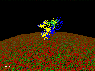
Attention: THIS PAGE IS UNDER CONSTRUCTION

Cellular Semiotics is a project for the Supercomputing '95 conference. The project is being developed at the National Center of Supercomputing Applications as a collaboration between COMPBIO Group, the illiView Group and Virtual Environment Group
Marcus Wagner mwagner@ncsa.uiuc.edu National Center for Supercomputing Applications (NCSA) Computational Biology University of Illinois at Urbana-Champaign 405 N. Mathews Av. Urbana, IL 61801
Alexei Bourd bourd@math.uiuc.edu NCSA and Department of Mathematics University of Illinois at Urbana-Champaign
Shankar Subramaniam shankar@ncsa.uiuc.edu NCSA and Department of Physiology and Biophysics University of Illinois at Urbana-Champaign
Eric Jakobsson jake@ncsa.uiuc.edu NCSA and Department of Physiology and Biophysics University of Illinois at Urbana-Champaign
George Francis gfrancis@mathiris.math.uiuc.edu Mathematics Department University of Illinois at Urbana-Champaign
Chris Hartman hartman@math.uiuc.edu Mathematics Department University of Illinois at Urbana-Champaign
Scott Banister banister@uiuc.edu Department of Computer Engineering University of Illinois at Urbana-Champaign
The project utilizes SGI Challenge Arrays and Convex Exemplar parallel supercomputers to compute Brownian dynamics using the University of Houston Brownian Dynamics (UHBD) program. The project uses Message Passing Interface (MPI) for the communication between supercomputers and the CAVE.
Here is scenario of the project
The scene begins with a picture of human anatomy (from a physiology and anatomy text or illustrations) with rotations and zooming in and out. The commentary could begin with introduction to human physiology.
Here you will see one aspect of human physiology at work. Every instant the human body is invaded by zillions of organisms (bacteria and viruses) and through a mechanism that has evolved over millenia the human body combats these and destroys the invaders. Occasionally, the body either lags behind in the combat (suffers temporary losses) or rarely loses the battle gradually. In such occasions, the human body is diseased from the microbial invasion. The physiological system in the human body that fights invasion is called the immune system. The immune system has numerous functions. First, it has to recognize the invader; second, it has to communicate to machinery that an invader has been located; third, it has to specifically localize the invader; fourth, it has to signal the heavy artillery to attack the invader and fifth, it has to ensure that the attack is limited only to the invader and finally, it has to memorize the invader's strategy to arm itself for future attacks.
The immune system also has to be versatile. It should fight microbes, it has never seen before in its evolutionary saga. Like every organized armed forces, it should have methods for reconnaissance and for targeting and attacking. The way the human body does this is to diversify the immune system into two parts - the humoral immune system: which produces immunoglobins or antibodies, that identify, bind specifically (localize) and escort the invader to the destruction machinery. These are called B-cells. -the cellular immune system: which regulates the build up of the attack, provides the heavy artillery and completes the attack process. These are called T-cells. The B-cells produce antibodies that float in the human serum and keeps constant surveillance of invaders.
At this stage we should show a close up of the blood flow system and focus on the serum. For this, we can have floating cells which have antibodies sticking out of them moving in the serum in a random manner. I like George's suggestion of having the sound effect of the heart beat. The graphics requires the flow system, caricature of antibody cells, their motion and perhaps other proteins of cell-bodies floating.
Let us now focus our attention on a B-cell. Its life begins in the bone marrow and the antibodies it produces end up in the serum. This cell, like every other cell in the body, contains all genetic information about the human body. However, it has specialized in producing antibodies. Let us get a close-up of what this cell looks like.
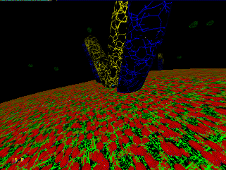
At this stage we should have a picture of a cell (from a physiology or cell biology textbook with all its contents highlighted. If it is desirable, we can have a commentary that provides background info. on all the organelles of the cell.
The cell wall also called the membrane is made up of lipid and protein molecules. The lipids have a dual personality in the sense that the insides are hydrophobic (water hating) and outsides are hydrophilic (water loving). This provides a leit motif for self-organization and membrane is a large sheet-like structure containing ordered lipid molecules in constant random and ordered motion.
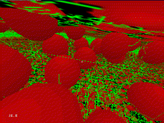
During this commentary, we show the membrane in different perspectives, inside, outside, top down, etc. When we talk of motion, we show a) periodic motion that Alexis has already got and superimpose on it one of See-Wing’s trajectories so that you can see some random motion. We can show water (how?) outside and none inside to highlight the dual behavior of the membrane.
The cell membranes also have proteins embedded in them. Some of the immunoglobins are first anchored on the B-cell, while several of the immunoglobin molecules are also released into the mileu around the cells. All of this accomplished Through complicated signaling pathways the cell has evolved.
We need to show here antibody molecules anchored on the membrane. I have a composite slide of an antibody molecule. Is it possible to scan it into the computer - that would be dramatic if possible. We can also then show motions of the antibody molecule.
The Y shaped molecules you see on the membrane are the antibody molecules. These are constantly interacting with scores of the microbes and biomolecules in the serum. All the particles in the medium are constantly undergoing random and directed motion while simultaneously interacting with every other particle around it. The random motion is very mush like the Brownian motion you see of dust particles in a medium. Antigens (those microbes or molecules that bind antibodies) are constantly diffusing and occasionally are steered towards the specific antibody molecules. The diffusional motion is so fast compared to some of the slower motions of the cell membranes that you see several thousands of collision events during the slow undulatory motion of the membrane.
Here we have the show of the membrane undulating and in faster timescale scores of flashes on the antibodies signaling the constant interactions and the timescales.
Once a specific antigen is recognized by the antibody molecule on a B-Cell, a complex signaling pathway is initiated which eventually ends in the cell producing a proliferation of antibody molecules. We will now walk through a choreographed mating between the antibody and the antigen.
Here we will witness the actual binding of the antibody to the antigen. Using supercomputers we will follow the diffusional motion of an antigen and we will see the steering effect of the antibody which will periodically result in the formation of a specific complex between the antibody and the antigen. The antibody you are seeing is an antibody specific for hen-egg lysozyme.
Here we give the antibody and we will walk the reader through the antibody structure and features/ Both general and things specific for D44.1 will be given. This is yet to be written.
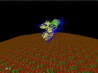
In what follows we will have real science where we will depict the docking of HEL onto D44.1 with BD and ES. Representationally, I suggest, we have ball stick models of the antibody (ribbons background ?) and show a dot surface for electrostatics that is in almost background. For HEL (lysozyme) we show again the ball and stick and color scheme according to change.
We will show trajectories and those that are successful and give a commentary on the events that happen. When the binding occurs we will signal the completion by having some graphical effect(any suggestions?).
Once the initial recognition and mating process has begun, proximal interatomic interactions take over and complete the tight binding. Here we show a close up of a molecular architecture and then turn on the dynamics. I recommend we do a 20 ps dynamics using quanta charmm and clock the trajectory and show it repeatedly for the close up. It is sufficiently random as to not appear periodic. This will produce a very nice effect.
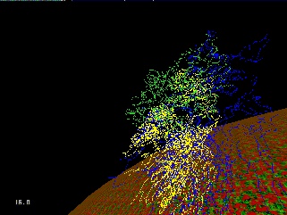
We can color code the atoms according to their change and we can show specific hydrogen bonds (dotted lines) of interactions. We can comment about the interatomic forces and their role.
Once the molecular mating is complete the signaling begins. The cell goes through birth pangs and generates hundreds of clones to the antibody that just mated with the antigen. In addition, the antigen-bound antibody call is target now for recognition by the T-cell which is the destruction machinery.
Here we will show only the clonal reproduction. We can zoom out of the molecular framework and go to the antigen bound antibody, zoom to the next level of antigen-bound-antibody-bound to a cell surface. That will complete one of the story we are telling.
Please e-mail to bourd@math.uiuc.edu with any suggestions.
Last Update: 9/26/95.